28 Jan Soft Tissue Ultrasound
Indications:
Skin and soft tissue infections: Assessing abscesses, cellulitis, and necrotizing fasciitis
Foreign body detection: Locating foreign objects embedded in soft tissue
Probe Choice:
Linear
Anatomy:
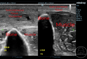
A side by side comparison of soft tissue affected by cellulitis with the pertinent structures: subcutaneous tissue, muscle, and bone. Check our vascular access / nerve block sections for anatomy related to vascular / nervous structures.
Pathology:
Abscess
Heterogenous fluid collection seen here. Primarily hypoechoic with mixed hyperechoic foci within (likely purulent material)
Pressing down with the probe causes this fluid to move, called the squish sign.
Cellulitis
Thickening of the soft tissue is seen here.
Cobblestoning: edema infiltrating the soft tissues which has the appearance of cobblestone paving of streets.
Necrotizing fasciitis
Soft tissue gas is seen here with the diffuse hyperechoic foci. It has dirty shadowing to it, which is gray in appearance, as compared to “clean” shadowing which is black or an
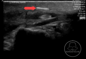
Foreign body
Foreign body appears as a hyperechoic structure in the soft tissue. They can have varied appearances with varied degrees of posterior shadowing, however, on the examination it is most likely going to be hyperechoic, or bright white.
Artifacts:
Posterior acoustic shadowing
Shadowing is seen deep to the bone. This is in contrast to the dirty shadowing of the necrotizing infection seen above.
Posterior acoustic enhancement
Tissues deep to a fluid filled cyst/abscess may appear brighter, this is termed posterior acoustic enhancement.
Pearls:
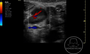
Color Doppler: When assessing fluid collection, use color doppler to evaluate for vasculature that you would otherwise not want to perform an incision and drainage on.
Septic thrombophlebitis seen above. Likely will not be on your examination, but demonstrates the importance of adding color doppler to ensure you are not cutting into a blood vessel.
Pitfalls:
Lymph nodes often mistaken for fluid collection/abscess
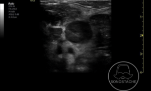
Here we see a lymph node with color flow, not to be mistaken for an abscess.