03 May Case of the week! 5/29/24 – Shoulder Pain
70 YOM presenting with shoulder pain
Scroll down for the answer!
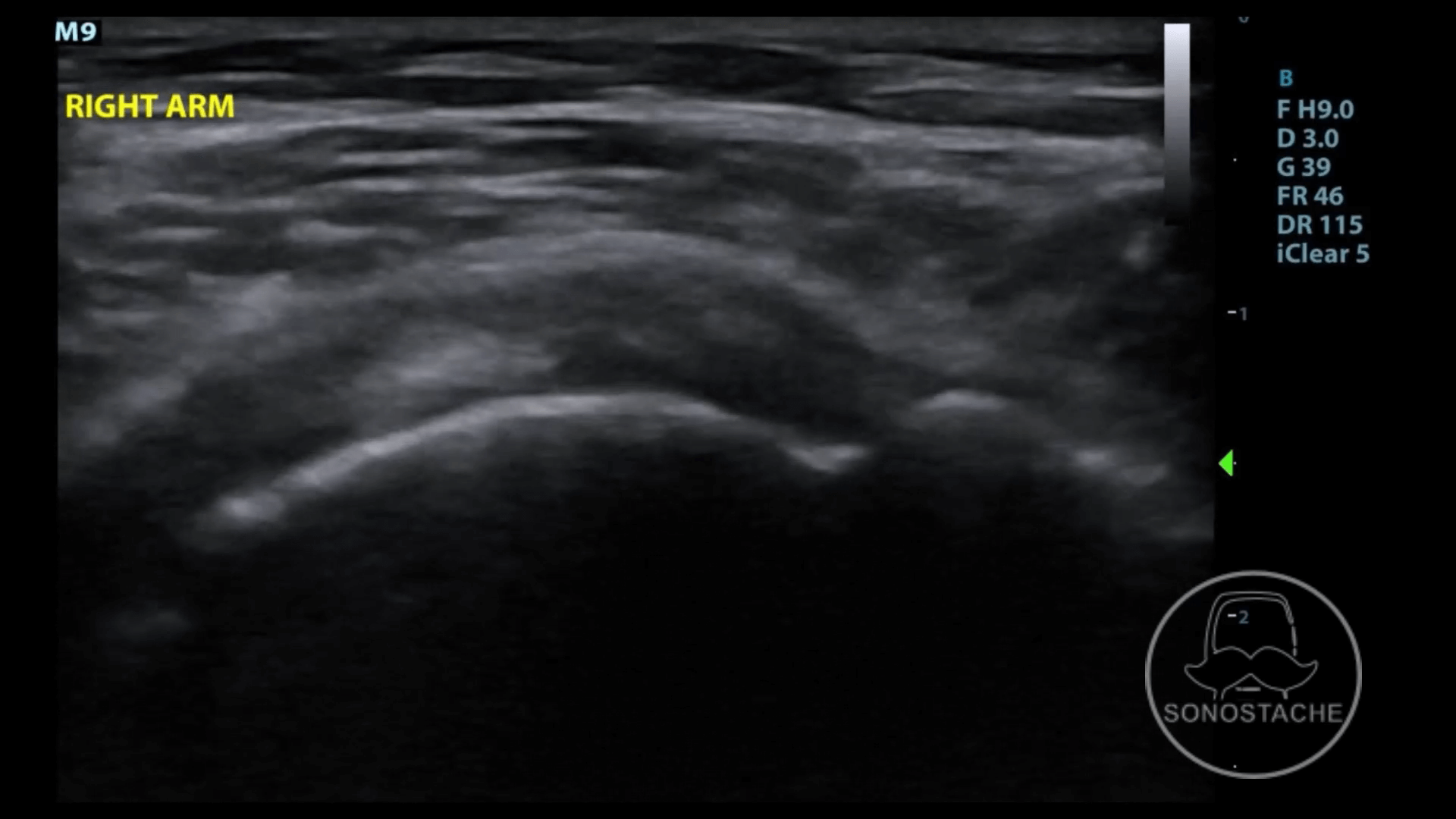
Answer: Supraspinatus tendinosis with partial tear
Loss of the homogenous appearance of healthy tendons are commonly seen with degenerative tendinosis. These can appear hypoechoic as fluid deposits between the tendon fibers. To complicate the picture, they are often concomitant with partial rotator cuff tears, and can be difficult to detect with ultrasound if the two overlap in the same area. With chronic tendinosis, calcium deposition may develop in the tendons and/or the subacromial-subdeltoid bursa. While it may have the classic appearance of smooth, well-circumscribed calcifications with posterior shadowing, they may also sometimes appear “fluffy” without shadowing. Ultrasound guided needle puncture and lavage has become a more common approach prior to surgical management.
Like any tendon, the supraspinatus can develop complete or partial thickness tears. The most common site is the anterior border. Tendon retraction is most indicative of full thickness tear, additional features can be noted including fluid in the subacromial-subdeltoid bursa or the glenohumeral joint itself.
Partial tears are more commonly noted with a focal discontinuity of the tendon. Ellman has classified partial thickness tears into grade 1 (less than 3mm), grade 2 (3-6mm) and grade 3 (>6mm). They can be further divided into low grade (<50% thickness) or high grade (>50% thickness). It is also common to see articular surface involvement, with a cortical bone irregularity of the greater tuberosity as a sensitive sign of partial thickness tear. A de-laminating tear occurs when the partial tear propagates proximally. A rim rent tear involves the articular surface near the footplate of the tendon. Large tears of the supraspinatus can also involve the infraspinatus, as both these tendon insertions overlap.
Ultrasound is a sensitive and specific tool for diagnosing supraspinatus tears (88%, 93%, respectively), but it is less reliable in distinguishing partial thickness and full thickness tears.
POCUS Pearl: Almost 90% of rotator cuff pathology occurs at the Critical Zone. The critical zone is the 1cm avascular area proximal to the greater tuberosity, so be sure to include this area with your scans!
Thanks!
-Sonostache Team
Follow us on X, @Sonostache
Follow us on Instagram, @Sonostache
Resources:
Singh JP. Shoulder ultrasound: What you need to know. Indian J Radiol Imaging. 2012 Oct;22(4):284-92. doi: 10.4103/0971-3026.111481.
Akhtar A, Richards J, Monga P. The biomechanics of the rotator cuff in health and disease – A narrative review. J Clin Orthop Trauma. 2021 Apr 26;18:150-156. doi: 10.1016/j.jcot.2021.04.019.
Guerini H, Fermand M, Godefroy D, Feydy A, Chevrot A, Morvan G, Gault N, Drapé JL. US appearance of partial-thickness supraspinatus tendon tears: Application of the string theory. Pictorial essay. J Ultrasound. 2012 Feb;15(1):7-15. doi: 10.1016/j.jus.2011.12.001. Epub 2012 Jan 15.