24 Apr Case of the week – 4/22/24 Cardiac Abnormality
What abnormality is seen?
Scroll down for the answer!
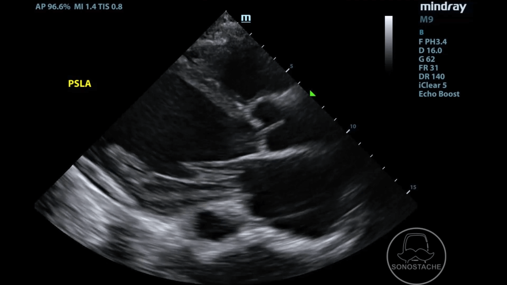
Answer: Dilated coronary sinus
The coronary sinus (CS) is a muscular tube that runs parallel to the atrioventricular junction. It runs adjacent to the left atrium, and is contained in a sheathe composed of atrial myocardium. CS dilation is often seen with dilated cardiomyopathy (also noted in this clip). CS size can also be increased in patients with CHF and pulmonary hypertension.
In the parasternal long axis, the CS is visualized posterior left atrioventricular junction, just above the descending aorta. As both are seen as circular structures, it can further be differentiated as the coronary sinus moves with cardiac motion as opposed to the descending aorta.
In the apical 4 chamber view, the transducer can be angled more posteriorly to visualize the coronary sinus in the atrioventricular groove. In the 2 chamber view, the coronary sinus can be seen at the atrioventricular junction to the left of the upper atrium as a small circle/oval.
Lastly, a prominent coronary sinus can be seen in persistent left superior vena cava. Persistent left superior vena cava is a rare congenital abnormality that is often draining into the coronary sinus.
Suspect coronary sinus with a round circular structure in the PSLAX or apical 2 chamber view that is contained within the pericardium, with normal diameter of 0.5cm to 0.9cm.
Thanks!
-Sonostache Team
Follow us on X, @Sonostache
Follow us on Instagram, @Sonostache
Resources:
Azizova, A., Onder, O., Arslan, S. et al. Persistent left superior vena cava: clinical importance and differential diagnoses. Insights Imaging 11, 110 (2020).
Son BJ, Kim H, Nam JH. Differential diagnosis for unusually dilated coronary sinus and right coronary artery incidentally found on echocardiography. J Yeungnam Med Sci. 2023;40(4):461-465.
Çakıcı M, Doğan A, Çetin M, Süner A, Polat M, Oylumlu M, Aktürk E, Abus S, Üçkardeş F. Coronary sinus dilatation is a sign of impaired right ventricular function in patients with heart failure. Anatol J Cardiol. 2015 Jul;15(7):542-7.