16 Apr Case of the Week – 4/15/2024, Adnexal Mass
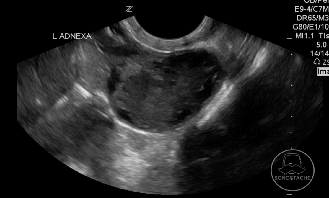
What abnormality is seen in this young, pregnant female?
Scroll down for the answer!
Answer: Corpus luteum cyst
Imaging and discerning features:
-One of the most common masses seen in the first trimester of pregnancy
-Secretes progesterone to support pregnancy until pregnancy takes over. Usually regresses by 16-18 weeks. Seen in 95% of pregnancies.
– < 5 cm in diameter (occasionally up to 10 cm)
-Commonly unilocular
-Irregular, or crenulated contour
-Due to the follicular tissue that comprises the outer walls of the corpus luteal cyst, there is enhanced circumferential blood flow (“ring of fire” on Doppler)
-If internal hemorrhage is present, there may be internal septations or echogenic debris
-Difficult to differentiate from a pathological cyst.
-Variable echogenicity
While commonly seen as cystic, if the walls of the corpus luteal cyst is thick or the fluid has internal hemorrhage, the cyst may appear as a solid mass. Color doppler here is helpful as the internal aspects of the cyst will not have uptake, with prominent peripheral vascularity. Due to the increased vascularity of the cyst, rupture (where you can see focal interruption of the cystic wall) can produce significant hemoperitoneum.
Part of the differential for this includes ectopic pregnancy. The tubal ring of ectopic pregnancy is typically more echogenic than the ovarian parenchyma that composes the lining of the corpus luteum cyst.
Lastly, with gentle manipulation of the ultrasound transducer you can determine if the mass is attached or independent of the ovary. If the ovary and mass appear attached it is more likely corpus luteal cyst. If the ovary and mass separate, it is more concerning for ectopic pregnancy.
Thanks!
-Sonostache Team
Follow us on X, @Sonostache
Follow us on Instagram, @Sonostache
Resources:
Bonde AA, Korngold EK, Foster BR, Fung AW, Sohaey R, Pettersson DR, Guimaraes AR, Coakley FV. Radiological appearances of corpus luteum cysts and their imaging mimics. Abdom Radiol (NY). 2016 Nov;41(11):2270-2282.
Swire MN, Castro-Aragon I, Levine D. Various sonographic appearances of the hemorrhagic corpus luteum cyst. Ultrasound Q. 2004 Jun;20(2):45-58.
Blaivas M, Lyon M. Reliability of adnexal mass mobility in distinguishing possible ectopic pregnancy from corpus luteum cysts. J Ultrasound Med. 2005 May;24(5):599-603.
Stein MW, Ricci ZJ, Novak L, Roberts JH, Koenigsberg M. Sonographic comparison of the tubal ring of ectopic pregnancy with the corpus luteum. J Ultrasound Med. 2004 Jan;23(1):57-62.