22 Aug Abdominal Aorta
Indications:
Abdominal / flank pain
Undifferentiated hypotension
Probe Choice:
Curvilinear probe
Technique, Views & Anatomy:
Technique: Use the curvilinear probe, starting midline just above the umbilicus (classically the most superficial part of the descending aorta) with the indicator to the patient’s right and scanning up and down in the short axis view, and then rotating the probe with the indicator towards the patient’s head to visualize the aorta in long axis.
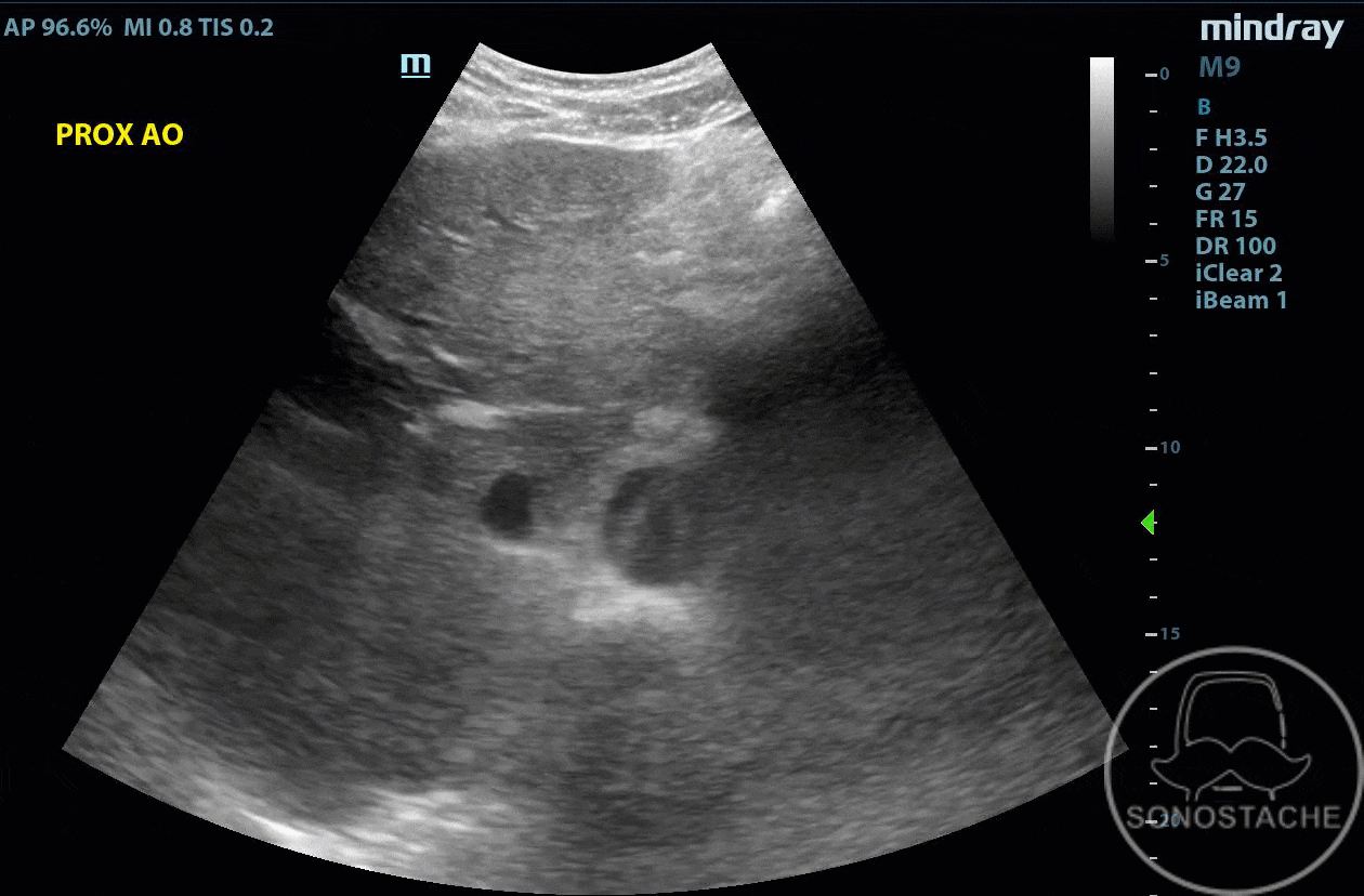
Visualize the aorta in short axis:
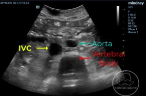
Identify the aorta, inferior vena cava, and the vertebral body. The vertebral body should be at the bottom of your image with shadowing, and is the landmark to find first as the aorta lies just above and to the right (on screen) above this structure.
When measuring the aorta, make sure to measure in the widest diameter, from outer wall to outer wall:
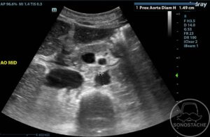
Measuring outer-to-outer will make sure you don’t accidentally exclude a thrombus, which should be included in measurement.
Visualize the aorta’s bifurcation into the common iliac arteries:
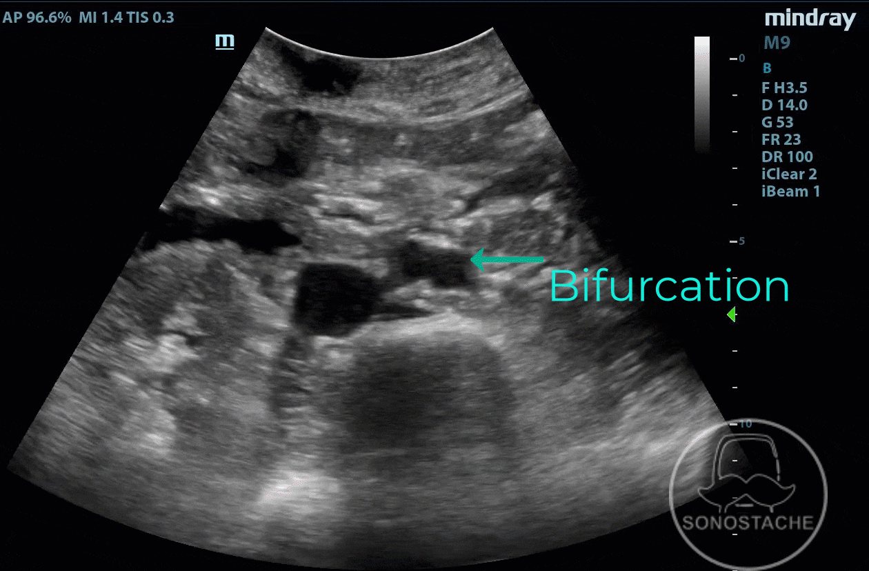
The majority of aortic aneurysms are infra-renal, so scanning through the bifurcation will help visualize these types of aneurysms.
Visualize the aorta in long axis:
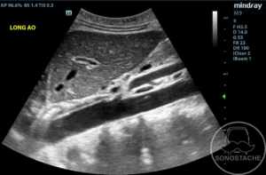
This last view of the aorta will help identify small saccular aneurysms that may not be found by clips of the aorta in short axis.
Pathology:
Abdominal Aortic Aneurysm (AAA):
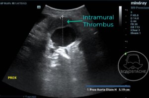
Enlarged aorta in short axis with mural thrombus, measuring 5.2cm.
Abdominal Aortic Dissection:
Abdominal aortic dissection seen in both long and short axis. As a clinical pearl, it is rare to have isolated abdominal aortic dissection. If you see one, suspect a thoracic dissection.
Artifacts:
Posterior Acoustic Shadowing – Clean or dirty?
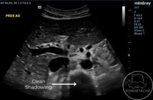
Here we see shadowing from the vertebral body. This is “clean shadowing” due to the absence of echos, dark shadowing. This is commonly seen with bone and dense gallstones.
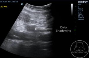
Here we see shadowing from bowel gas. This is “dirty shadowing” and looks grayer than the clean shadowing. This is considered normal when looking at structures that should have air (like bowel) but may be pathologic (like necrotizing fasciitis).
Pearls:
If bowel gas is obscuring your vision, apply gentle steady downward pressure while rocking the probe to maneuver the gas out of the way.
The maximum depth should be the spinal column. Often finding this structure first helps locate the aorta.
Pitfalls:
When measuring the aorta, measure outer wall to outer wall. This is because an intramural thrombus might cause a falsely low measurement, and should be included in the measurement.